by Dr. Robert Zepeki, DVM CVCP, owner of Village Animal Group
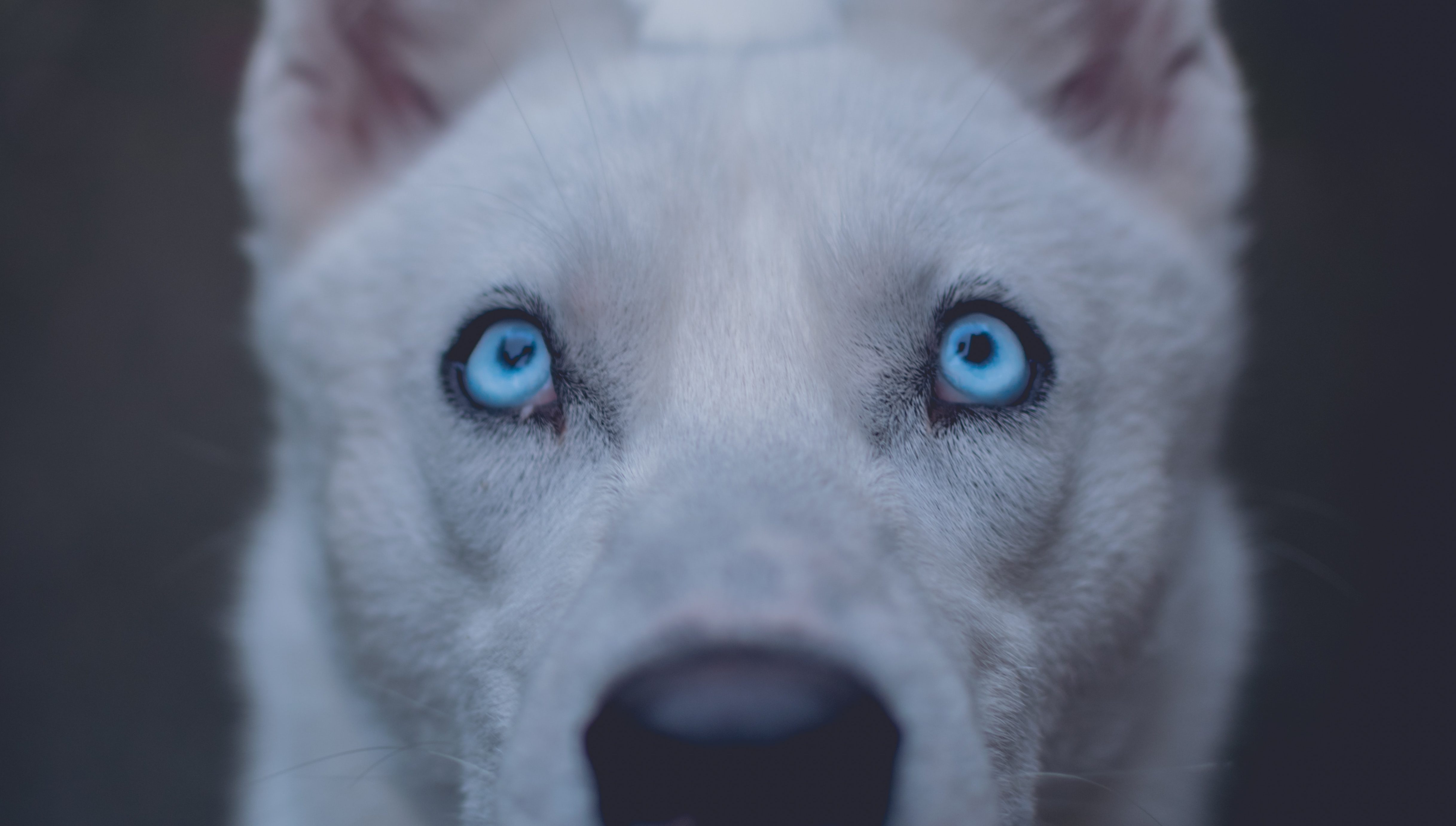
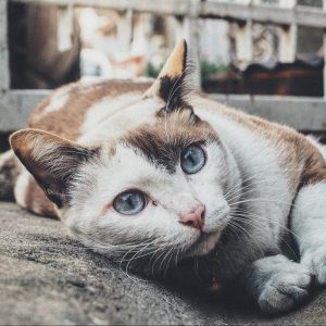
Your kitty and doggy Friends possess a remarkable stereoscopic vision. They inherited this ability from their hunting ancestors. Both species can see some color. Your doggy Friend has an eye very similar to our eye with one focal point on the back of the eye. However, the kitty Friends are endowed with very good night vision. Their eyes are so sensitive to light that the iris can close to a slit in strong light resulting in two focal points at the top and the bottom of the closed iris. You have probably noticed this when your kitty Friend sits in the sunlight. The eye consists of the following main structures; cornea, iris, lens, and globe or eyeball.
The Cornea
Cornea, or clear portion of the eye that you can see, is the clear top of a container that holds the clear liquid, the aqueous humor in the front chamber of the eye. The cornea consists of layers like an onion. There are normally no blood vessels running through the cornea. Increased pressure in the eye can cause the cornea to cloud by squeezing the layers together. The disease can easily attack the cornea from the outside or inside of the eye. Thorns, cat claws, doors, and cars are the most common sources of trauma to the eye in my experience. The tears that cover the cornea are a constant source of protection.
 If there are not enough tears, the cornea will begin to dry out. This is very serious. A cornea that has been injured or irritated for several days will begin to develop little red blood vessels coming in from the edge. This condition must receive immediate treatment. Scrapes or pits in the cornea are called corneal ulcers. These are tears in one or more layers of the cornea. Repair is very slow. These injuries must be medicated until they completely heal.
Sometimes the healing is not complete and a scar will result. The scar is usually white. This is simply an area where there is no living corneal tissue. Your friend can see around scars if they are not very large. If all of the layers of the cornea are penetrated down to the last layer, called Descemet’s Membrane, your friend is in immediate danger of having the eye collapse. Fortunately, you veterinarian can perform a surgery to close the eyelids over the cornea and allow it to repair.
The Lens
The lens is suspended behind the iris (that colored part of your friend’s eye). There are little elastic fibers that hold the lens in perfect alignment so that images that come through the small opening in the iris will be clearly projected to the retina. Your friends have lenses very similar in shape and structure to our human eye lenses. Trauma can cause one or more of these ligaments to come loose, causing the lens to drop or tilt. This could change the image on the back of the eye.
The lens becomes cloudy as your friend’s age. This is called nuclear sclerosis and causes a small loss of vision. Kind of like the steam on your windshield. The lens can also develop a cataract. This is a bright white spot. There are many types of cataracts that can develop for many reasons. Many breeds of dogs have a high incidence of cataracts. Diabetic friends can develop cataracts also. Fortunately, veterinary ophthalmologists can remove cataractous lenses as routinely as our human counterparts.
The Ciliary Body
The tissue in the corner formed by the iris with the front chamber of the eye (anterior chamber) is where the clear liquid in the eye is produced. This area is called the ciliary body. This system of cells continuously produces a very clear liquid that flows into the front chamber of the eye. There is also a drainage point at this place in the eye where the liquid leaves the chamber. There is a pressure control system in place to prevent an abnormal increase or decrease in the pressure in this chamber caused by the presence of this fluid. If the pressure is too low, the front chamber will deflate like a balloon. This condition usually occurs from trauma.
The Iris
 The iris (uvea) is a circular bundle of strands of muscles, the center of which is that tiny hole (aperture), through which the light image is projected to the retina through the lens. These muscles can be damaged, become infected or inflamed (uveitis) and swell, bleed or stick to the lens (iris bombe). Much of the pain in the eye is located in these muscles. Iris color is hereditary. If the iris sticks to the lens (iris bombe), your veterinary ophthalmologist can unstick it. The iris is a very delicate system. Fear can cause a dilation of the iris. Certain medicines can also dilate or constrict the iris. This is known as the ciliary body. It is a ring of muscles in various colors that open and close to let in light through the center of this ring.
Glaucoma
Increased pressure in the eye is much more damaging and can lead to blindness. The increased pressure can come from either too much fluid or a plugged drainage system. When the iris swells from inflammation or infection, the normal angle at which it attaches to the eye will partially or completely close the drainage system causing a pressure increase (glaucoma). Inflammation of the cells that secrete the fluid can cause an overload of fluid also causing an increase in pressure.
The aging process can cause sagging of the iris and the eye with a partial closure of this drainage system. This type of angle-closure glaucoma is quite common in dogs as they age but rarely occurs in cats. Glaucoma can be treated with some success. Currently, veterinary ophthalmologists can partially freeze the tissues that produce the aqueous humor, that clear liquid in the front of the eye, and reduce the pressure significantly.
Petworks has mobile vets on call if you need immediate care.
About the Author:
 Dr. Bob Zepecki brings experience, compassion, and innovation to All Pet Center in Hot Springs Village, Arkansas. His involvement in veterinary medicine has spanned all areas of applied practice and preventative medicine from exotic species such as lions and tigers, to domestic animals and pets.